The Role of Genetic Mutations in Melanoma Development

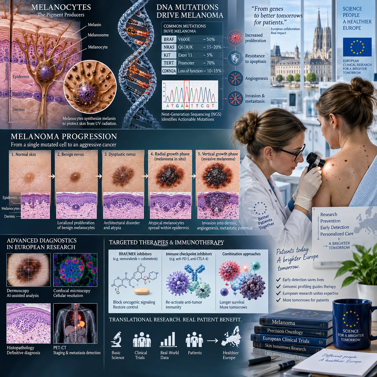
Melanoma develops when melanocytes, the pigment-producing cells of the skin and other tissues, acquire biological changes that allow uncontrolled growth and survival. Genetic mutations are central to this process, but no single mutation explains every melanoma. The disease is genetically diverse, and its development reflects interactions among DNA damage, inherited susceptibility, cellular regulation, immune responses, and environmental exposure.
Understanding these changes helps researchers classify melanoma and helps clinicians select appropriate tests or treatments for some patients. However, mutation status is interpreted alongside histopathology, tumour stage, imaging, clinical features, and multidisciplinary assessment.
How melanoma develops
Melanoma develops when melanocytes accumulate DNA changes that disrupt normal controls on cell division, repair, and survival. These changes can create a clone of abnormal cells that grows within the skin and may eventually invade surrounding tissue or spread to distant organs.
Healthy melanocytes respond to signals that regulate when they divide, pause, repair DNA, or undergo programmed cell death. Genetic mutations can interfere with these safeguards in several ways. Some mutations activate growth-promoting signals continuously; others disable tumour-suppressor proteins that normally restrain cell division or remove damaged cells.
Melanoma progression usually involves more than one biological alteration. An early mutation may give a melanocyte a growth advantage, while later changes help the tumour invade tissue, evade immune surveillance, or resist treatment. The order and combination of these events differ between tumours.
This helps explain why melanomas that look similar under a microscope can behave differently. A mutation is best understood as part of a changing cellular system rather than as a standalone diagnosis. Tumour location, depth, ulceration, lymph-node involvement, metastatic pattern, and immune activity also influence clinical behaviour.
Inherited versus acquired genetic mutations
Inherited, or germline, mutations are present in many cells from birth, whereas acquired, or somatic, mutations arise in individual cells during a person’s lifetime. This distinction matters because the two types of mutation have different implications for family risk, testing, and treatment.
A germline mutation can increase a person’s susceptibility to melanoma. One recognised example involves CDKN2A, a tumour-suppressor gene associated with familial melanoma in some families. A pathogenic germline variant does not guarantee that melanoma will develop, and many people with melanoma have no identifiable inherited mutation.
Somatic mutations are found only in tumour cells or in a subset of cells within a tumour. They are not usually passed to children. Ultraviolet radiation, errors during DNA replication, oxidative stress, and other cellular processes can contribute to these acquired changes. A tumour sample may therefore contain mutations that are highly relevant to treatment without indicating inherited cancer risk.
Genetic testing requires careful interpretation. Testing tumour tissue answers a different question from testing blood or saliva for germline variants. Germline testing may be considered when personal or family history suggests inherited susceptibility, while tumour profiling is commonly used to identify molecular features relevant to advanced disease or research. Results should be discussed with qualified healthcare professionals, and genetic counselling may be appropriate when an inherited risk is suspected.
Key genes and pathways involved in melanoma
BRAF, NRAS, NF1, and CDKN2A are among the genes frequently discussed in melanoma biology, while the MAPK pathway is a major growth-signalling network affected in many tumours. These genes describe important patterns, but they do not represent every melanoma and should not be treated as interchangeable markers.
BRAF and the MAPK pathway
The MAPK pathway transmits signals that influence cell growth and division. In some melanomas, an acquired change in BRAF keeps this pathway abnormally active. BRAF alterations, particularly activating variants, can therefore provide tumour cells with persistent growth signals.
When a clinically relevant BRAF alteration is identified, it may support consideration of BRAF-directed treatment combined with inhibition of a downstream MEK protein. Such therapy is designed around the biology of the tumour, but responses vary. Resistance can emerge through pathway reactivation, alternative signalling, changes within tumour-cell populations, or interactions with the surrounding tumour environment.
NRAS, NF1, and CDKN2A
NRAS is another signalling gene that can activate growth pathways, including MAPK-related signalling. NRAS-mutant melanoma is biologically distinct from BRAF-mutant disease, and the presence of an NRAS alteration does not automatically point to the same treatment strategy.
NF1 helps regulate signalling proteins. Loss-of-function changes in NF1 can contribute to abnormal pathway activity and are found in a subset of melanomas, including tumours that do not carry common BRAF or NRAS alterations. These tumours may be grouped as having a different molecular profile, although the category remains biologically diverse.
CDKN2A encodes proteins involved in cell-cycle control. When its tumour-suppressor function is lost, cells may progress through the cell cycle despite damage or abnormal growth signals. CDKN2A alterations can be acquired in a tumour or, less commonly, inherited as part of familial susceptibility.
Melanoma biology also involves other genes and mechanisms, including alterations affecting the cell cycle, telomere maintenance, chromatin regulation, and immune interactions. The key clinical lesson is that a mutation profile provides useful context, not a complete account of tumour behaviour.
How environmental and cellular factors contribute to mutation
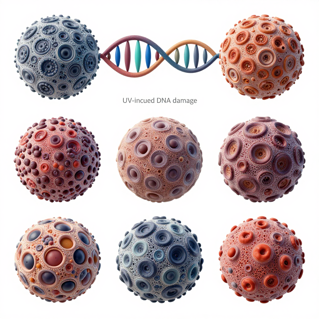
Ultraviolet radiation is an important source of DNA damage linked to melanoma, but melanoma development is multifactorial. UV-related injury can create characteristic DNA changes, particularly in melanocytes exposed to sunlight or artificial UV sources, yet exposure history alone does not predict every tumour’s genetic profile.
UV radiation can damage DNA directly and can also generate reactive molecules that affect cellular structures. Cells repair much of this injury, but unrepaired damage may become a permanent mutation when the cell divides. Repeated exposure, intense intermittent exposure, skin type, number and type of naevi, immune status, and inherited susceptibility can all influence risk.
Cellular context matters as well. A mutation may have different effects depending on which melanocyte contains it, whether nearby cells provide growth signals, and how effectively the immune system recognises abnormal cells. The tumour microenvironment includes immune cells, blood vessels, connective tissue, and signalling molecules that can shape progression.
These relationships make prevention messages more nuanced than a simple one-cause model. Reducing avoidable UV exposure remains relevant to skin health, but it cannot eliminate all melanoma risk. New lesions or changing lesions should be assessed by an appropriate healthcare professional rather than interpreted through genetic information alone.
From mutation profiling to diagnosis and treatment
Molecular profiling can identify tumour alterations that support classification, treatment selection, and research, but it does not replace pathology or clinical staging. Testing usually examines DNA, and sometimes RNA or protein activity, from a biopsy or surgical specimen.
Pathologists first establish whether a lesion is melanoma and describe features such as tumour thickness, ulceration, and margins. In advanced disease, molecular testing may then assess alterations such as BRAF, NRAS, NF1, or other clinically relevant markers. The choice of test depends on the disease setting, available therapies, laboratory standards, and the clinical question.
Results can influence two broad treatment approaches:
- Targeted therapy uses drugs designed to interfere with a specific altered pathway, such as BRAF and MEK signalling in selected BRAF-mutant melanomas.
- Immunotherapy stimulates or restores anti-tumour immune activity and can work across several molecular subtypes, although response is not guaranteed.
Mutation testing also supports trials investigating treatment combinations, resistance mechanisms, minimal residual disease, and tumour evolution. A single biopsy provides a snapshot. Metastases can contain different subclones, and the molecular profile may change after treatment. This is one reason research groups, including European collaborative networks such as the European Organisation for Research and Treatment of Cancer, continue to study longitudinal samples and real-world treatment outcomes.
What genetic mutations mean for risk, prognosis, and research
Genetic mutations can inform melanoma risk assessment and treatment planning, but they do not determine an individual outcome by themselves. Prognosis depends on the interaction of tumour genetics with stage, anatomical site, tumour burden, immune response, treatment response, and overall clinical circumstances.
An inherited CDKN2A variant may prompt more detailed family-risk assessment, whereas a BRAF mutation found in tumour tissue usually describes the cancer’s acquired biology. Confusing these findings can cause unnecessary concern among relatives or create false reassurance for people without a detected germline variant.
Mutation profiles also have limits. A test may fail to detect a relevant alteration because of low tumour content, technical constraints, tumour heterogeneity, or incomplete knowledge. Conversely, finding a mutation does not prove that it initiated the cancer or predict exactly how a person will respond to treatment.
A useful way to interpret molecular information is the three-context model:
- Cellular context: Which genes and pathways are altered, and how do they affect growth or immune escape?
- Tumour context: Where is the melanoma, how advanced is it, and are different subclones present?
- Clinical context: Which treatments, trials, comorbidities, and patient preferences shape the decision?
European melanoma research continues to refine this model through collaborative trials, molecular datasets, translational studies, and investigations into resistance. The aim is to match treatment more precisely while recognising that melanoma remains a heterogeneous disease. Patients and families should ask their clinical team whether a result is somatic or germline, what action it supports, and which uncertainties remain.
Frequently asked questions about melanoma mutations
Are all melanoma mutations inherited?
No. Most mutations found in a melanoma are acquired somatic mutations within tumour cells. A smaller proportion of people carry inherited variants that increase susceptibility, and tumour testing cannot automatically determine whether a mutation is germline.
Which genetic mutations are commonly found in melanoma?
BRAF, NRAS, and NF1 are important acquired mutation categories, while CDKN2A can be altered in tumours and can also be involved in inherited familial melanoma susceptibility. The exact profile varies by tumour and patient population.
Does having a BRAF mutation cause melanoma?
A BRAF mutation can contribute to melanoma development by activating growth signalling, but it is not the sole cause of melanoma. Other genetic, cellular, environmental, and immune factors are usually involved.
How is melanoma mutation testing performed?
Testing generally uses DNA extracted from a melanoma biopsy or surgical specimen. Blood or saliva testing is used when clinicians are investigating possible inherited susceptibility. The method and genes examined depend on the clinical purpose.
Can genetic mutations affect melanoma treatment?
Yes. Some tumour mutations can identify patients who may be eligible for targeted therapy or clinical trials. Immunotherapy may also be considered across multiple molecular subtypes. Treatment decisions still require pathology, staging, medical history, and specialist assessment.
Genetic mutations provide a powerful window into melanoma development, but they are one part of a larger clinical picture. Research-led interpretation helps turn molecular findings into safer, more relevant decisions for patients.